The Elusive Neurotrophic Keratitis
The cornea is the first line of defense of the eye, and as such is the most densely innervated and sensitive tissue in the body. With a central corneal nerve density of approximately 7,000 nociceptors per square millimeter the cornea is 300 to 600 times more sensitive than skin.1 These nerves play a vital role in protecting the cornea and maintaining and preserving ocular health and vision. But when these nerves become compromised or impaired, corneal sensation decreases leading to neurotrophic keratitis.
Neurotrophic keratitis is a rare, potentially sight-threatening, degenerative, corneal disease caused by damage anywhere along the pathway of the fifth cranial nerve: from the Trigeminal nucleus to the corneal nerve endings.2 This damage causes a loss of corneal sensation which leads to tear film abnormalities, epithelial breakdown, impairment of healing, ulceration and in severe cases corneal melting and perforation.3
Although the most common cause of neurotrophic keratitis is infection, specifically herpetic keratitis (varicella zoster or herpes simplex), several conditions can lead to impairment of trigeminal innervation of the cornea.4 They can be classified as:
 Ocular:
• Infection (herpes)
• LASIK/PRK
• PK/DALK
• Chemical or Physical burns
• Chronic contact lens overwear
• Topical drop toxicity (especially those containing benzalkonium chloride)
• Chronic ocular surface injury
 SYSTEMIC:
- Diabetes
- Multiple Sclerosis
- Vitamin A deficiency
- Amyloidosis
- Drugs (antipsychotics, antihistamines, neuroepileptic drugs)
- Leprosy
 CENTRAL NERVOUS SYSTEM:
• Stroke
• Post-neurosurgical procedures
• Neoplasm
• Aneurysms
• Degenerative disorders of the CNS
 GENETIC:
• Riley-Day syndrome
• Goldenhar-Gorlin syndrome
• Mobius syndrome
• Familial corneal hypoesthesia
Regardless of the source, patients with neurotrophic keratitis rarely complain of pain. This makes early diagnosis extremely challenging, as patients may not seek medical advice until the condition has progressed to a more advanced stage.
Since treatment for neurotrophic keratitis is based on the severity of the disease, Mackie proposed a classification system that divides the condition into three stages.5
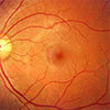
Stage 1 is considered mild neurotrophic keratitis and is characterized by decreased tear breakup time, punctate epithelial erosion, and corneal edema. Superficial neovascularization and stromal scarring may also be present in eyes with long-standing mild NK.

Cornea Fluorescein staining in mild NK (source: Transitional Vision Science and Technology: Assessment of Corneal Fluorescein Staining.2019;Vol. 8; No.6; Article 34)
Stage 2 represents moderate disease and the hallmark of this stage is the persistent or recurrent epithelial defect. It is usually located inferiorly with smooth, rolled edges. There may be loose, edematous epithelium surrounding the defect with stromal edema and Descemet’s membrane folds. With long-standing disease, there can also be anterior chamber inflammation.

Neurotrophic corneal ulcer in moderate NK (source: Canadian Journal of Ophthalmology: Topical Insulin for Neurotrophic Corneal Ulcers.May 14, 2020)
Stage 3 represents severe neurotrophic keratitis with corneal ulceration. The ulcer can progress to stromal melting and eventually to corneal perforation.

Corneal ulcer that has progressed to stromal melting in severe NK (© 2021 American Academy of Ophthalmology)
The primary goal of treatment for neurotrophic keratitis is to restore corneal integrity through stimulating epithelial re-growth and to prevent progression of the disease. Patients should use preservative-free eye medications and avoid topical NSAIDs as these can further decrease corneal sensitivity. Ocular surface diseases other than those associated with neurotrophic keratitis such as blepharitis, meibomian gland dysfunction and dry eyes should be properly treated.
The following table shows a categorized treatment approach to therapy options that are available for neurotrophic keratitis. Treatment varies depending on the stage of the disease and its severity and these are not mutually exclusive.4,6

Despite the various medical and surgical therapies proposed, neurotrophic keratitis remains one of the most difficult and challenging ocular diseases encountered in clinical practice. To effectively manage these patients, prompt identification and early treatment are critical since the prognosis largely depends on the severity and duration of the disease when treatment is initiated. With careful attention to the many facets of neurotrophic keratitis, we can facilitate corneal healing, maintain ocular integrity and ultimately preserve vision.
References:
- Yang A, Chow J, Liu J. (2018) Corneal Innervation and Sensation: The Eye and Beyond. Yale J Biology and Medicine. 2018;91(1): 13–21.
- Mastropasqua L, Massaro-Giordano G, Nubile M, Sacchetti M. Understanding the pathogenesis of neurotrophic keratitis: the role of corneal nerves. J Cell Physiology. 2017;232(4): 717-724.
- Semeraro F, Forbice E, Romano V, Angi M, Romano MR, Filippelli ME, Di Iorio R, Costagliola C. Neurotrophic keratitis. Ophthalmologica. 2014;231(4):191-7.
- Dua H, Said D, Messmer E, et al. Neurotrophic keratopathy. Prog Retin Eye Res. 2018;66:107-131.
- Mackie IE. Neuroparalytic keratitis. In: Fraunfelder F, Roy FH, Meyer SM, eds. Current Ocular Therapy. WB Saunders; 1995:452-454.
- Sacchetti M, Lambiase A. Diagnosis and management of neurotrophic keratitis.Clin Ophthalmol. 2014;8:571-579.

Author: Kerri Svanda, OD
Specialties: Medical Eye Care
Seattle
The cornea is our window to the world. Keeping it clear and healthy can sometimes be a long and challenging endeavor. I chose this topic after recently co-managing two very difficult cases of neurotrophic keratitis following cornea and cataract surgeries. As part of the Seattle cornea team with Dr. Audrey Talley Rostov and Dr. Stacey Keppol, I am honored to work with our referring doctors to help our patients achieve their best possible vision, be it through cornea surgery, cataract surgery or refractive surgery.