During a recent retinal consultation, I encountered a case in which the end result was the diagnosis of a large choroidal melanoma. The past ocular history was reviewed and was significant for a “suspicious choroidal nevus” identified approximately 1.5 years ago by a local optometrist. Unfortunately, the patient did not return for follow-up to his optometrist until he had a recent change in vision. Currently, the medical workup and treatment decisions have yet to be made for this patient and the final outcome is pending. Keeping in mind that the 5 year survival rate of small choroidal melanoma versus large melanoma is 84% vs 47%, respectively. In cases like this, I often wonder what could have been done to identify this lesion as an early malignancy and how the outcome could have been altered if caught sooner.
The evaluation of suspicious pigmented choroidal lesions are a daily requirement in the life of a retinal specialist. This assessment includes the use of multiple diagnostic tools (or multimodal imaging) in conjunction with the clinical exam to determine the diagnosis or in some cases, provide a relative degree of malignant suspicion. Twenty years ago, we relied heavily upon clinical characteristics, supplemented by the diagnostic tools of BScan imaging and fluorescein angiography to make decisions. However, over the past two decades there have been significant advancements in diagnostic technologies which fortunately can be employed in assisting with the identification of choroidal melanoma. These technologies can allow us to determine malignancies earlier and hopefully have an impact in the long term outcomes for these patients.
To quote Carol Shields, MD at Wills Eye Hospital in Philadelphia, the goal is to, “Find small ocular melanoma.” Her collaborative work is paramount in the diagnosis and treatment of these tumors and she recently provided an update to her approach to identification. She has upgraded her classic pneumonic to TFSOM-DIM. This translates, To Find Small Ocular Melanoma, Do IMaging. This is a reminder to identify the following significant characteristics: Thickness greater than 2mm (BScan or OCT), subretinal Fluid (OCT), Symptoms, Orange pigment (fundus autofluorescence), Melanoma acoustic hollowness (BScan) and Imaging (fundus photos showing lesions greater than 5 mm in diameter and absence of drusen.) The real world application of implementing imaging technologies can greatly assist the eyecare provider in making the correct diagnosis. This can be illustrated when evaluating the case mentioned above in the following actual examples:
Figure 1: OCT demonstrating SRF and “shaggy photoreceptors” finding
Figure 2: OCT demonstrating SRF and hyperreflective deposits correlating to orange pigment
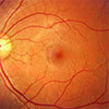
Figure 3: Fundus photo demonstrating lesion greater than 5mm in diameter, no drusen
Figure 4: Fundus autofluorescence demonstrating spotty fluorescence of orange pigment
Figure 5: BScan demonstrating acoustic hollowness, thickness greater than 2mm and RPE excavation
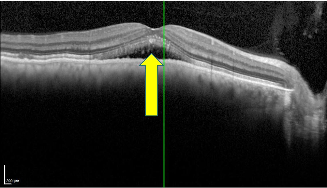

Figure 1 Figure 2

Figure 3


Figure 4 Figure 5
As delineated by the world’s experts, the early identification of ocular melanoma can be greatly assisted by imaging. In many of our clinical practices, we have several of these technologies that can be utilized to image suspicious choroidal lesions to recognize their potential for malignancy. Implementing these diagnostics in the care of these patients will allow eyecare providers to raise the bar in their ability to identify small ocular melanomas at earlier stages which should lead to better long term patient outcomes.

Richard Lee, OD, FAAO
Specialty: Medical Eye Care
My topic of identifying choroidal melanoma stems from a desire to inform community colleagues of the updated algorithms to improve patient care. Early identification is critical when caring for these patients.
As a provider at NW Eye Surgeons, I enjoy caring for all patients with vitreoretinal disorders and collaborating with community providers. It is my passion to provide the best care available in the state of Washington to all patients who entrust us with their medical needs.
References
Eye cancer – statistics. Cancer.Net. (2020, February 14). Retrieved November 13, 2022, from https://www.cancer.net/cancer-types/eye-cancer/statistics#:~:text=The%205%2Dyear%20relative%20survival%20rate%20for%20people%20with%20small,large%20choroidal%20melanoma%20is%2047%25. Shields, C. L., Lally, S. E., Dalvin, L. A., Sagoo, M. S., Pellegrini, M., Kaliki, S., Gündüz, A. K., Furuta, M., Mruthyunjaya, P., Fung, A. T., Duker, J. S., Selig, S. M., Yaghy, A., Ferenczy, S. R., Eydelman, M. B., & Blumenkranz, M. S. (2021). White Paper on ophthalmic imaging for Choroidal Nevus Identification and transformation into melanoma. Translational Vision Science & Technology, 10(2), 24. https://doi.org/10.1167/tvst.10.2.24