
Cornea,Cornea Transplant,Provider Newsletter
 In 1904 Edward Zirm performed the first successful full thickness corneal transplant in a person. It is reported that the person went on to see well enough to work for a number of months after his surgery. For about 100 years we have performed penetrating keratoplasty and restoring patients’ vision. Everyone would like to improve our outcomes and do a better more successful surgery when possible. Several great leaders have pioneered the field of cornea in the last 20 years.
There have been several attempts at selective replacement of corneal tissue with the most successful being from Garett Melles from the Netherlands. In 1998, he described selectively replacing just Descemet’s membrane and the endothelium to help patients who only suffered from endothelial decompensation – Fuchs’ corneal dystrophy and pseudophakic corneal edema. He called this Descemets Membrane Endothelial Keratoplasty or DMEK. Roughly half of the transplants performed in the US are for this indication.
Before this procedure became popular other surgeons had attempts at evolving partial thickness transplants to selectively replace the endothelium while maintaining the anterior cornea and ocular surface. Several main struggles with the tried and true full thickness penetrating keratoplasty are related to the large circular wound held together with sutures. Irregular astigmatism is a significant problem that limits full visual rehabilitation after a full thickness transplant (penetrating keratoplasty). Many patients cannot get crisp 20/20 vision with glasses and are not able to insert and remove a hard contact lens that could provide the crisp quality 20/20 vision. This means that after a full thickness transplant, the full potential vision is not realized. Complications such as wound dehiscence and suture abscess are also a significant limitation to full thickness transplantation. Drs. Busin, Gorovoy, Melles, Price and Terry all pioneered deep lamellar keratoplasty and what we now term Descemet’s Stripping (Automated) Endothelial Keratoplasty or DSEK (DSAEK). The automated means the lamellar graft is prepared with a microkeratome instead of by hand dissection. Essentially everywhere a microkeratome is available, the microkeratome is used and it is automated. The terms are often used interchangeably. DSEK evolved into the procedure we now know from about 2003 to 2006. The advantages were significant – more rapid healing without irregular astigmatism and fewer complications due to minimal or no sutures and smaller incisions. DSEK’s rapid adoption by surgeons was dramatic due to its significant advantages over PKP performing 1,398 DSEK in 2005 and 17,468 cases in 2008. In 2011-2012 the number of endothelial keratoplasty cases comprised over 50% of all transplants performed.
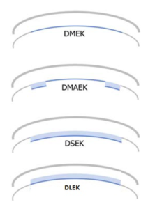
DMEK was originally a technically difficult procedure to perform due to the thin graft thickness and outward coiling of the endothelial cells relative to the Descemet’s membrane. There were several versions of grafts between DSEK and DMEK called DMAEK and DMEK-S but these while these were easier to perform inside the eye their donor preparation was significantly more challenging. Near or after 2010, the techniques for performing DMEK had improved and its popularity has increased. Remember it was described in 1998 but it took more than 10 years before many surgeons were able to perform the technique and it was becoming more popular with more surgeons. DMEK has the same advantages over full thickness transplants as DSEK but also has advantages over DSEK in better visual acuity and lower rejection rates. It appears that the less tissue transplanted the lower the rejection rate meaning that in general terms that full thickness transplant has the highest rates of rejection, followed by DSEK followed by thin DSEK followed by DMEK. Visual acuity is great with DSEK but even better with DMEK.
A new form of corneal surgery that challenges our thinking of endothelial cells is Descemet’s stripping only (DSO) or Descemetorhexis without Endothelial Keratoplasty (DWEK). Why is this radical? We were classically taught that endothelial cells do not divide or replicate. While this may still be true, endothelial cells it seems can at least spread out and move around if they have space and are encouraged. DSO or DWEK simply strips away the central 4 mm of endothelial cells and then does not replace the bare stroma with any transplant tissue. Some patients, possibly with the assistance of certain eye drops called Rho kinase inhibitors, can heal and have perfect vision WITHOUT any chance of rejection. This form of surgery is currently being studied to understand who makes the best candidate and how to improve the results of successful surgery.
Another new and exciting form of corneal surgery on the horizon for the same disease type is from cell based therapy. Endothelial dysfunction such as Fuchs’ dystrophy and pseudophakic corneal edema may be treated by a simple injection of cells or with stripping away the central pathology and then a small needle injection of cells into the eye. The thought of and possibility of essentially performing a corneal transplant by injecting cells through a needle is totally different from anything available currently and continues the rapid evolution in the field of corneal transplantation. It is an exciting time to be a corneal surgeon, and also better to be a patient in need of these safer and better surgeries!

Michael R. Banitt, MD, MHA
Smokey Point Clinic
I chose this topic because I am passionate about the cornea and cornea transplants. I want to always provide the best practice and outcomes for my patients. To be able to provide the best surgery and outcomes for my patients means always changing with current trends. In the case of cornea transplants this has been a rapid evolution with the need to adapt quickly to these new surgeries and the need to continue to evolve to maintain the best outcomes in the future.

