
Provider Newsletter,Thyroid Eye Disease
Thyroid eye disease is an autoimmune condition affecting the ocular tissues. It occurs in up to 25% of patients with Graves’ disease. It is most common in patients who have hyperthyroidism (Graves’ disease), however, it can also occur under eurthyroid (10%) or autoimmune mediated hypothyroid conditions. Thyroid eye disease is more common in women, typically around 30 years old, but with another peak near 60 years of age. Men, older individuals, and smokers tend to develop more severe disease.
The most common course is triggered by the immune system recognizing the thyroid stimulating hormone receptor (TSHR) as an antigen. This triggers an inflammatory cascade mediated by T-cells and B-cells that results in fibroblast and adipocyte proliferation through activation of TSHR and connected insulin like growth factor (IGF-1) receptors. These receptors may also be upregulated in retro-orbital tissues of patients with thyroid eye disease. Hydrophilic glycosaminoglycans such as hyaluronic acid are produced and lead to swelling of soft tissues.
At the time of diagnosis approximately 20% of patients will have normal thyroid levels without any history of abnormal test results, 40% will have hyperthyroidism, and 20% will develop hyperthyroidism within 6 months of developing the ocular signs and symptoms. The rest develop ocular complications following treatment of hyperthyroidism. Hypothyroidism following treatment with radioactive iodine for hyperthyroidism is a risk factor for development of thyroid eye disease.
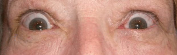
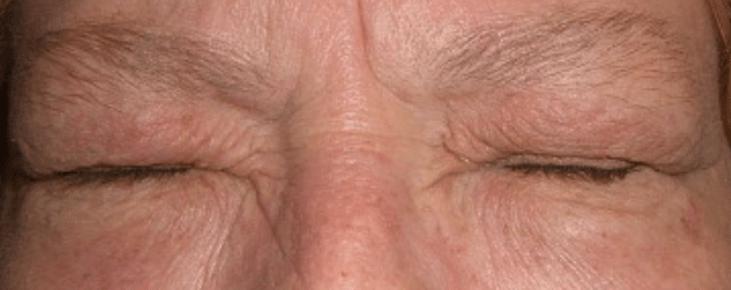
Diagnosis of thyroid orbitopathy is made based on the presence of swelling of eyelids (image 2) or conjunctiva, eye pain, eye pain with eye movement, presence of conjunctival injection or eyelid erythema, and inflammation of the plica or caruncle. If more than 3 of these findings are present, then there is active thyroid eye disease (see table 1). Severity grading is based on the amount of lid retraction, exophthalmos, soft tissue involvement, corneal involvement, diplopia, and optic nerve involvement (see table 2). Lid retraction (image 1) and lid lag (upper lids remain elevated on downgaze) may be signs of hyperthyroidism and may improve with normalization of thyroid hormone levels.
Table 1: Clinical Activity Score (CAS)
| Initial exam: add up positive findings on items 1-7 |
| 1) Spontaneous orbital pain or pressure |
| 2) Gaze evoked orbital pain or pressure |
| 3) Eyelid edema-if considered to be secondary to active Graves’ orbitopathy |
| 4) Eyelid erythema |
| 5) Conjunctival injection – if considered to be secondary to active Graves’ orbitopathy |
| 6) Chemosis |
| 7) Inflammation of caruncle or plica |
| Follow-up exam (1-3 months): add up positive findings on items 8-10 |
| 8) Increase of >2mm proptosis |
| 9) Decrease in ocular excursion in any direction of >8 degrees |
| 10) Decrease in acuity equivalent to 1 Snellen Line |
| Active orbitopathy exists if score is >3/7 on initial exam or >4/10 on follow-up |
Table 2: Severity Chart for Graves Orbitopathy
| Characteristic | Mild | Moderate to Severe | Sight Threatening |
| Eyelid retraction | <2mm | >/=2mm | |
| Exophthalmos Norms by race & gender Asian (Thai) F/M: 16/17 (18.6 Chinese) Black F/M: 23/24 White F/M: 19/21 Norm is </= 2mm between eyes | <3mm from norm | >/=3mm from norm | |
| Soft-tissue involvement | Mild | Moderate to severe | |
| Diplopia | None to transient | Inconstant to constant | |
| Corneal involvement | Absent to mild | Moderate to severe | Severe |
| Optic nerve involvement | None | None | Compression |
Symptoms of hyperthyroidism include rapid heart rate, heat intolerance, anxiety, excessive sweating, difficulty sleeping, and tremors. Patients may also exhibit other manifestations of Graves’ disease such as enlarged thyroid gland, pretibial myxedema (red, swollen skin of lower leg), or acropachy (swollen skin on hands and clubbing of fingers). If diagnosis is uncertain, or in the setting of unilateral or severe orbitopathy, consider CT or MRI orbits without contrast to evaluate for swelling of extraocular muscles and proptosis. Never use iodinated contrast as it may exacerbate hyperthyroidism or interfere with radioactive iodine therapy.
Labs to consider are: TSH, total T3 (triiodothyronine), free T4 (thyroxine), TRAb (Thyroid stimulating receptor antibodies: thyroid stimulating immunoglobulin (TSI) & TSH receptor binding inhibitory antibodies (TBII)), Tg (thyroglobulin), and TgAb (thyroglobulin antibodies), and TPOAb (thyroid peroxidase antibodies). Thyroglobulin and thyroid peroxidase antibodies can be found in those without thyroid disorders and in those with Hashimoto’s thyroiditis as well. In hyperthyroidism, TSH will be low, T3 and T4 will usually be high. Graves orbitopathy is more likely to occur in those with TRAb and TgAb. The course of the disease is likely to be more severe if the TSI is above 400. Approximately 5% of patients develop a severe course of the disease. Thyroid stimulating immunoglobulin will often elevate following radioactive iodine therapy. The levels peak approximately 4 months following therapy and may remain elevated for years. The clinical activity score (Table 1) is used to measure response to therapy.
Therapy consists of treatment to normalize thyroid levels. Methimazole, radioactive iodine, and/or thyroid surgery are used to reduce hyperthyroidism and levothyroxine is used to treat hypothyroidism. Levothyroxine should be used promptly following radioactive iodine therapy. Glucocorticoids should be used concurrently with radioactive iodine if the patient is at increased risk for orbitopathy. Methimazole is typically used for 1-2 years. Therapy also consists of methods to control symptoms such as a beta blocker to reduce rapid heart rate, shaking, nervousness, and anxiety; and corticosteroids are used to reduce swelling and inflammation.
The course of the eye disease is somewhat independent of treatment of thyroid hormone levels and is typically active for 1 to 3 years. Untreated hyperthyroidism can exacerbate orbitopathy but treated hyperthyroidism does not reduce the risk of orbitopathy. Moderate to severe orbitopathy is a contraindication to radioactive iodine therapy. Orbitopathy treatment is based on severity and activity. Counsel patients on the importance of smoking cessation to lessen risk of more severe orbitopathy. Use lubricating drops and anti-inflammatory drops to control dry eye. Consider sleep goggles if exposure is a concern. Sleep with the head of the bed elevated to minimize edema. Use eye pressure lowering drops to control ocular hypertension or glaucoma. Consider patching or prism glasses to minimize symptoms from diplopia.
Mild disease may improve with 100mcg Selenium twice daily by mouth, however, long term Selenium has been associated with a higher risk of type 2 diabetes. Moderate to severe disease in the active phase typically will respond to immunomodulating therapy. Prednisone or methylprednisolone are primary treatment options. Prednisone is used at 30mg/d for 4 weeks, then tapered to the lowest daily dose for maintenance of symptoms. Methylprednisolone is used for more severe orbitopathy, or if not responding to oral prednisone. It is dosed through IV at 500mg per week for 6 weeks, followed by 250mg weekly for the next 6 weeks. If severe optic nerve compression is present, the patient should be admitted to the hospital for 4g IV methylprednisolone and possible orbital decompression surgery. Teprotumumab is a newer treatment option for patients not responding to steroid therapy. It is administered by IV. The first two doses are given over 90 minutes but if tolerated well, the next 6 doses can be administered over 1 hour. It is dosed every 3 weeks for 8 doses. The first dose is 10mg/kg, the rest are 20mg/kg. Mycophenolate mofetil or tocilizumab may be other steroid sparing immunomodulatory therapies effective at reducing orbitopathy symptoms. Surgical intervention via orbital decompression or external orbital radiation may be necessary for sight threatening orbitopathy. Orbital decompression surgery is sometimes used for cosmesis once a patient has reached an inactive phase. Other surgeries to consider in the inactive phase are strabismus surgery to reduce diplopia and eyelid surgery to reduce eyelid retraction. Strabismus surgery should be performed after orbital decompression.
Thyroid eye disease can cause serious limitations on a patient’s quality of life. As primary eye care providers, we may be the first to diagnose a patient with this condition and help get them the treatment that they need. Prompt diagnosis and treatment will help maintain vision and improve their quality of life.
References:
Davies, T.F. (2021) Pathogens of Graves’. In J.E. Mulder (Ed). UpToDate. Retrieved on 07/20/21 from https://www.uptodate.com/contents/pathogenesis-of-graves-disease?search=graves&source=search_result&selectedTitle=2~150&usage_type=default&display_rank=2
Davies, T.F., Burch, H.B. (2021) Treatment of Graves’ orbitopathy (ophthalmopathy). In J.E. Mulder (Ed). UpToDate. Retrieved on 07/20/21 from https://www.uptodate.com/contents/treatment-of-graves-orbitopathy-ophthalmopathy?search=thyroid%20eye%20disease&source=search_result&selectedTitle=2~62&usage_type=default&display_rank=2
Ross, D.S. (2021) Graves’ hyperthyroidism in nonpregnant adults: Overview of treatment. In J.E. Mulder (Ed). UpToDate. Retrieved on 07/20/2021 from
Sathyadeepak, R. Thyroid Eye Disease: Its Causes and Diagnosis. Review of Ophthalmology. 2018 Nov 9. Retrieved 07/20/21 from https://www.reviewofophthalmology.com/article/thyroid-eye-disease-its-causes-and-diagnosis
Gupta, L, Prendes M.A., Douglas, R. (2021) Teprotumumab. In J. Giacometti (Ed). UpToDate/EyeWiki. Retrieved on 07/20/21 from https://eyewiki.aao.org/Teprotumumab
Ponto KA, Kanitz M, Olivo PD, Pitz S, Pfeiffer N, Kahaly GJ. Clinical relevance of thyroid-stimulating immunoglobulins in graves’ ophthalmopathy. Ophthalmology. 2011 Nov;118(11):2279-85. doi: 10.1016/j.ophtha.2011.03.030. PMID: 21684605.
Lytton SD, Ponto KA, Kanitz M, Matheis N, Kohn LD, Kahaly GJ. A novel thyroid stimulating immunoglobulin bioassay is a functional indicator of activity and severity of Graves’ orbitopathy. J Clin Endocrinol Metab. 2010 May;95(5):2123-31. doi: 10.1210/jc.2009-2470. Epub 2010 Mar 17. PMID: 20237164.

Author: Davina S. Kuhnline, OD
Specialties: Medical Eye Care
I chose thyroid eye disease because it is a topic that I am interested in and wanted to find more information on and I thought it may be useful for others as well. I learned the NO SPECS classification in school and found the CAS table more useful. I enjoy working with you to help manage challenging and unusual cases.

